توضیحات
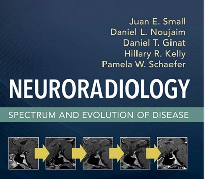
Neuroradiology Spectrum
size: 186 MB
SECTION I
Brain
Parenchymal Hemorrhage and Trauma, 1
۱٫ Brain Parenchymal Hematoma Evolution, 1
Juan E. Small
۲٫ Subdural Hemorrhage and Posttraumatic
Hygroma, 6
Lindstly A.N. Duy llftd Jutm E. Smail
Disorders of Cerebral Vascular
Autoregulation, 20
۳٫ Posterior Reversible Encephalopathy Syndrome, 20
Girisb Br~thkl tmtJ 1Jrmw Policmi
Arteriopathy, 32
۴٫ Cerebral Amyloid Angiopathy, 32
Mimi!Umst
Metabolic Disorders, 41
S. Wernicke Encephalopathy, 41
AmvnB. Paul
۶٫ Central Pontine Myelinolysis, 46
Juan E. S’INIII, Dtmiel L. Noujaim, tmtJ Arll)(J 0. Blllkeb
Infection, 50
۷٫ Herpes Simplex Encephalitis, 50
Gme M. ffiimtein tmtJ Jutm E. Smail
۸٫ Toxoplasmosis, 56
Ytm Setm Xie llftd ]11S0111UmtJwerlter
۹٫ N eurocysticercosis, 61
Samir Noujllim, Ltnormce Baborm~, Dtmiel L. Noujllim,
II1Jd Marie Tumimla
Autoimmune/Inflammatory Disorders, 80
۱۰٫ Acute Disseminated Encephalomyelitis, 80
Girish Br~thkl II1Jd 1Jrmw Policmi
۱۱٫ Autoimmune Encephalitis, 96
Ftmg Frtmlr Yu II1Jd Jutm E. Smail
۱۲٫ Progressive Multifocal Leukoencephalopathy, 103
AmvnB. Paul
۱۳٫ Central Nervous System-Immune Reconstitution
Inflammatory Syndrome, 108
AmvnB. Paul
X
۱۴٫ Neurosarcoidosis, 115
Girisb B11tbkl, Ptmltllj Wllflll, rmd Brtmo Policmi
Tumors, 136
۱ S. Treated Gliomas, 13 6
Ftmg Fnmle Yu rmd Om! Rapa/i7w
۱۶٫ Hemangioblastoma, 153
Dtmiel L. Noujllim rmd Jadyn A. Therrien
Ventricular System Alterations, 158
۱۷٫ Intracranial Hypotension, 158
Dr~eHee Kim rmd Jutm E. Smail
۱۸٫ Idiopathic Intracranial Hypertension
(Pseudotumor Cerebri), 163
Dr~eHee Kim llftd Jutm E. Smail
Pituitary Abnormalities, 167
۱۹٫ Partially Empty Sella, 167
JeJfny A. HMbim tmd Jutm E. Smail
۲۰٫ Rathke Cleft Cyst, 172
JeJfny A. HMbim tmd Seyed &zapour
۲۱٫ Pituitary Apoplexy, 181
Dtmiel L. Noujaim
Neurodegenerative Disease, 186
۲۲٫ Hypertrophic Olivary Degeneration, 186
Jutm E. Smail
SECTION II
Spine
Degenerative Disease, 189
۲۳٫ Ossification of the Posterior Longitudinal
Ligament, 189
Niltbtmiel Temin, Mmn~ Galper, II1Jd Jutm E. Small
۲۴٫ Lumbar Interbody Fusion, 199
Dll’lm Martin II1Jd Jutm E. Smail
Posttraumatic Effects, 208
۲S. Kummel Disease, 208
Dtmiel L. Noujaim
Infection, 213
۲۶٫ Discitis-Osteomyelitis, 213
Adam P. Brytmt, Dtmiel L. Noujr~im, II1Jd Toshio Morittmi
۲۷٫ Tuberculous Spinal Infection, 222
Adam P. Bryant tmd Tosbio Mrlrittmi
Bone Lesions, 226
۲۸٫ Chordoma, 226
LoW Go/Jm tmd Jutm E. Small
۲۹٫ Vertebral Hemangioma, 232
Mimi!Umst
Cord Lesions, 236
۳۰٫ Syringohydromyelia, 236
AlmmB.RnJ
۳۱٫ Spinal Cord Infarction, 241
Mimi K1mst, Dtmn Mimin, tmd Vutor Hugo Perez Perez
۳۲٫ Subacute Progressive Ascending Myelopathy, 250
SamaAlrh11’111 tmd Jutm E. Smtdl
Cord Tumors, 254
۳۳٫ Spinal Cord Ependymoma, 254
Omar RJruez tmd WdlimnA. Mebtm Jr.
۳۴٫ Spinal Cord Astrocytoma, 266
Jutm E. Small
Spine Deformity, 277
۳۵٫ Hirayama Disease, 277
Jutm E. SmtJIJ, Doreen T. Ho, tmd Dtnm Martin
۳۶٫ Dorsal Thoracic Arachnoid Abnormalities, 282
Dtmiel L. Nrmjflim, Dtmn Mimin, Walter L. CbflmfJirm,
tmd Juan E. Small
SECTION Ill
Head and Neck
Infection, 291
۳۷٫ Orbital Infection, 291
Daniel T. Ginat
۳۸٫ Recurrent Suppurative Thyroiditis Due to
Piriform Sinus Fistula, 298
Daniel T. Ginat tmd Jutm E. Small
Contents xi
Inflammatory Disorders, 302
۳۹٫ Thyroid-Associated Orbitopathy, . 302
Pauley Cbea, Emily Rut~m, Philip D. &usoubris,
tmd SuzamJe K Freitag
۴۰٫ IgG4-Related Disease in the Head and Neck, 308
Detm T. Jiffery tmd Hillary R. KJJy
۴۱٫ Sjogren Syndrome, 318
DlmielLAm tmd Dtmiel T. Ginat
۴۲٫ Cholesteatoma, 322
RnJ M. Brmcb tmd Hillllry R. KElly
۴۳٫ Labyrinthitis, 331
RnJ M. Brmcb tmd Hillllry R. KElly
Tumors, 339
۴۴٫ Paraganglioma, 339
KAtherine L. Reimbagm tmd Hillllry R. &lly
۴۵٫ Esthesioneuroblastoma, 346
RnJ M. Brmcb tmd Hillllry R. KElly
Posttraumatic Effects, 357
۴۶٫ Vocal Cord Augmentation/Injection
Laryngoplasty, 357
Daniel T. Ginat, Juan E. SmtJIJ, tmd Dtmiel L. Noujaim
Bone Lesions, 363
۴۷٫ Otospongiosis, 363
KAtherine L. Reimhagm tmd Hillllry R. &lly
۴۸٫ Paget’s Disease, 3 68
Juan E. Smtdl
Vascular Lesions, 381
۴۹٫ Carotid Blowout Syndrome, 381
Daniel T. Ginat tmd Juan E. Small
This page intentionally left blank
INTRODUCTION
Magnetic resonance imaging (MR1) can differentiate between acute,
subacute, and chronic hemorrhage because of its sensitivity and
specificity to hemoglobin degradation products. Therefore the
imaging interpreter is, with proper knowledge, able to estimate
the age of a brain parenchymal hematoma. The blood products in
a hematoma evolve through a predictable variation in hemoglobin
oxygenation states and hemoglobin byproducts. This predictable
pattern of hematoma evolution over time leads to a specific pattern
of changing signal intensities on conventional MRI.
There are limitations to the accuracy of hematoma age interpretation.
Several direct and indirect factors, including the operating
field strength of the magnet, the mode of image acquisition, and
a wide range of biologic factors particular to the patient, may
affect the imaging evolution of a parenchymal hematoma. Despite
substantial variability, it is generally accepted that five stages of
parenchymal hemorrhage can be distinguished by MRI. A basic
understanding of the biochemical evolution of brain parenchymal
hemorrhage and magnetic properties that affect MRI signal are
essential for interpretation.


